Parathyroid gland sonography is a critical diagnostic tool in the field of endocrinology and radiology. This non invading imaging technique plays a polar role in the espial and evaluation of parathyroid disorders, which can importantly impingement calcium metamorphosis and overall health. Understanding the intricacies of parathyroid gland sonography is crucial for healthcare professionals take to furnish accurate diagnoses and effective treatments.
Understanding the Parathyroid Glands
The parathyroid glands are small-scale, pea size glands located behind the thyroid gland in the neck. Typically, there are four parathyroid glands, although the figure can vary. These glands create parathyroid hormone (PTH), which regulates calcium and phosphorus levels in the body. Dysfunction of the parathyroid glands can lead to conditions such as hyperparathyroidism and hypoparathyroidism, both of which require precise diagnostic methods for accurate management.
The Importance of Parathyroid Gland Sonography
Parathyroid gland sonography, also known as parathyroid ultrasound, is a valuable diagnostic creature that uses eminent frequency sound waves to make images of the parathyroid glands. This visualise technique is specially utile in place unnatural parathyroid glands, such as those that are enlarged or have develop tumors. The master advantages of parathyroid gland sonography include:
- Non invading nature, making it a safe and comfy procedure for patients.
- Ability to detect parathyroid adenomas, which are benign tumors that can make hyperparathyroidism.
- Usefulness in point fine needle aspiration biopsies and other interventional procedures.
- Cost effectiveness equate to other imaging modalities like CT scans and MRI.
Preparation for Parathyroid Gland Sonography
Preparing for a parathyroid gland sonography procedure is relatively straightforward. Patients are typically apprize to:
- Wear comfy cloak that allows easy access to the neck area.
- Avoid wearing jewelry or other accessories that might interfere with the fancy summons.
- Inform the healthcare provider about any allergies or medical conditions that could involve the function.
No special dietetical restrictions are ordinarily required, but it is essential to postdate any specific instructions provided by the healthcare provider.
The Parathyroid Gland Sonography Procedure
The parathyroid gland sonography subprogram is loosely quick and painless. Here is a step by step overview of what to expect:
- The patient is positioned comfortably on an examination table, usually lying on their back with the neck slightly extended.
- A h2o ground gel is use to the neck country to enhance the transmittance of sound waves.
- The sonographer uses a transducer, a handheld device that emits and receives sound waves, to scan the neck region.
- The transducer is moved mildly over the skin, and the sound waves create existent time images of the parathyroid glands and surrounding structures.
- The images are displayed on a monitor and can be bewitch for further analysis.
The entire routine typically takes about 15 30 minutes.
Note: Patients may experience slight discomfort from the pressing of the transducer, but the subroutine is generally well endure.
Interpreting Parathyroid Gland Sonography Results
Interpreting the results of parathyroid gland sonography requires expertise in radiology and endocrinology. The images incur during the subprogram are canvass for several key features:
- Size and shape of the parathyroid glands.
- Presence of any nodules or masses.
- Location of the parathyroid glands comparative to the thyroid gland.
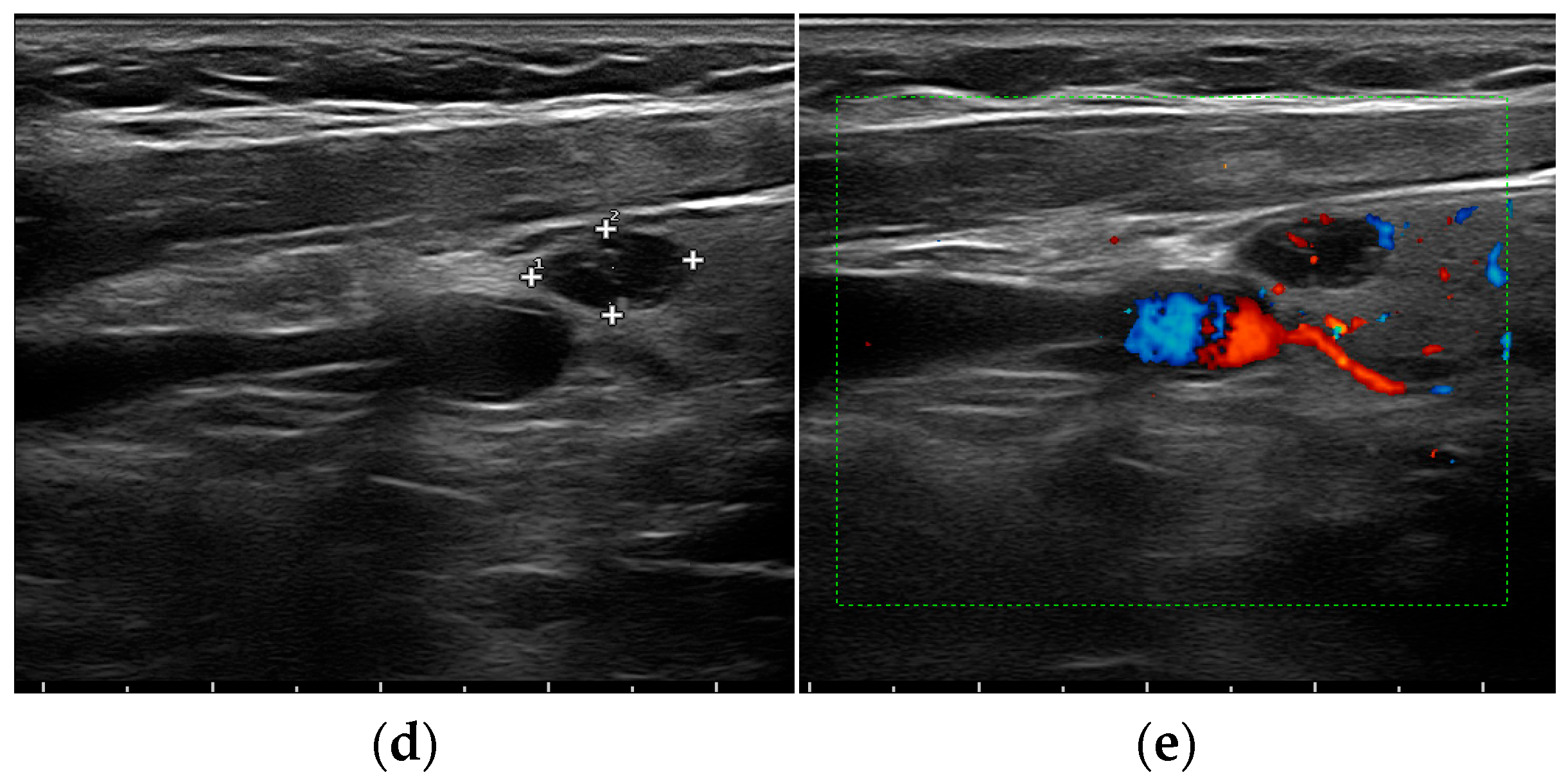
- Blood flow patterns within the glands, which can show the presence of tumors.
Abnormal findings may include enlarge parathyroid glands, the presence of adenomas, or other structural abnormalities. These findings are all-important for diagnose conditions such as chief hyperparathyroidism, secondary hyperparathyroidism, and parathyroid carcinoma.
Common Findings in Parathyroid Gland Sonography
Parathyroid gland sonography can expose various findings that are indicatory of different parathyroid disorders. Some of the most common findings include:
| Finding | Description | Potential Diagnosis |
|---|---|---|
| Enlarged Parathyroid Glands | Glands that are larger than normal. | Hyperparathyroidism |
| Parathyroid Adenomas | Benign tumors within the parathyroid glands. | Primary Hyperparathyroidism |
| Parathyroid Carcinoma | Malignant tumors within the parathyroid glands. | Parathyroid Carcinoma |
| Ectopic Parathyroid Glands | Glands located outside their normal position. | Hyperparathyroidism |
Limitations of Parathyroid Gland Sonography
While parathyroid gland sonography is a valuable diagnostic tool, it does have certain limitations. These include:
- Difficulty in visualizing small or deeply place parathyroid glands.
- Limited power to differentiate between benign and malignant tumors.
- Potential for false negative results, especially in cases of ectopic parathyroid glands.
- Dependence on the skill and experience of the sonographer.
In some cases, additional imaging modalities such as CT scans, MRI, or nuclear medicine studies may be take to complement the findings of parathyroid gland sonography.
Advanced Techniques in Parathyroid Gland Sonography
Advances in medical technology have led to the development of more sophisticated techniques in parathyroid gland sonography. Some of these progress methods include:
- Doppler Ultrasound: This technique uses sound waves to visualize blood flow within the parathyroid glands, aid to identify hypervascular tumors.
- Elastography: This method assesses the stiffness of the parathyroid glands, which can show the front of tumors or other abnormalities.
- 3D Ultrasound: This provides a more detail and comprehensive view of the parathyroid glands, aid in the detection of small-scale or ectopic glands.
These advanced techniques enhance the diagnostic accuracy of parathyroid gland sonography and improve patient outcomes.
Role of Parathyroid Gland Sonography in Surgical Planning
Parathyroid gland sonography plays a crucial role in operative plan for patients with parathyroid disorders. Preoperative imaging helps surgeons to:
- Locate the abnormal parathyroid glands accurately.
- Plan the operative approach, whether it be a minimally invading parathyroidectomy or a more extensive procedure.
- Identify any potential complications, such as ectopic glands or vascular anomalies.
Accurate preoperative project can importantly reduce operative time, belittle complications, and improve patient recovery.
Post Procedure Care and Follow Up
After undergo parathyroid gland sonography, patients typically do not require any especial post procedure care. However, follow up appointments may be necessary to discuss the results and plan further management. Patients should:
- Follow any instructions cater by the healthcare supplier affect follow up care.
- Report any unusual symptoms or concerns to their healthcare provider.
- Attend all scheduled follow up appointments to admonisher their condition and adjust treatment as needed.
Regular follow up is crucial for deal parathyroid disorders and ensuring optimal health outcomes.
Note: Patients with unnatural parathyroid gland sonography results may require additional symptomatic tests or interventions, such as fine needle dream biopsies or surgical procedures.
Parathyroid gland sonography is an indispensable creature in the diagnosis and management of parathyroid disorders. Its non invading nature, cost effectiveness, and ability to ply detailed images make it a choose choice for many healthcare professionals. By translate the intricacies of this imaging technique, healthcare providers can proffer more accurate diagnoses and efficacious treatments, ultimately ameliorate patient outcomes. The integrating of advanced techniques and the role of parathyroid gland sonography in surgical design further enhance its value in modern medical practice.
Related Terms:
- parathyroid gland ultrasound appearances
- parathyroid gland ultrasound radiology
- parathyroid tumor ultrasound
- parathyroid adenoma ultrasound
- parathyroid ultrasound normal
- parathyroid placement ultrasound