The humerus is the long bone in the arm that runs from the shoulder to the elbow. It plays a crucial role in the movement and constancy of the upper limb. Understanding the humerus bone markings is all-important for aesculapian professionals, anatomists, and students of human anatomy. These markings function as attachment points for muscles, ligaments, and tendons, and they provide worthful information about the bone's function and construction.
Anatomy of the Humerus
The humerus is dissever into several distinct regions, each with its own set of humerus bone markings. These regions include the head, neck, body, and distal end. The head of the humerus articulates with the glenoid cavity of the scapula to form the shoulder joint. The body, or shaft, of the humerus is the long, cylindrical portion that extends from the neck to the distal end. The distal end includes the sidelong and median epicondyles, the trochlea, and the capitulum, which articulate with the bones of the forearm to form the elbow joint.
Proximal Humerus Bone Markings
The proximal end of the humerus features several important humerus bone markings that are important for understanding the bone s mapping and the muscles that attach to it. These markings include:
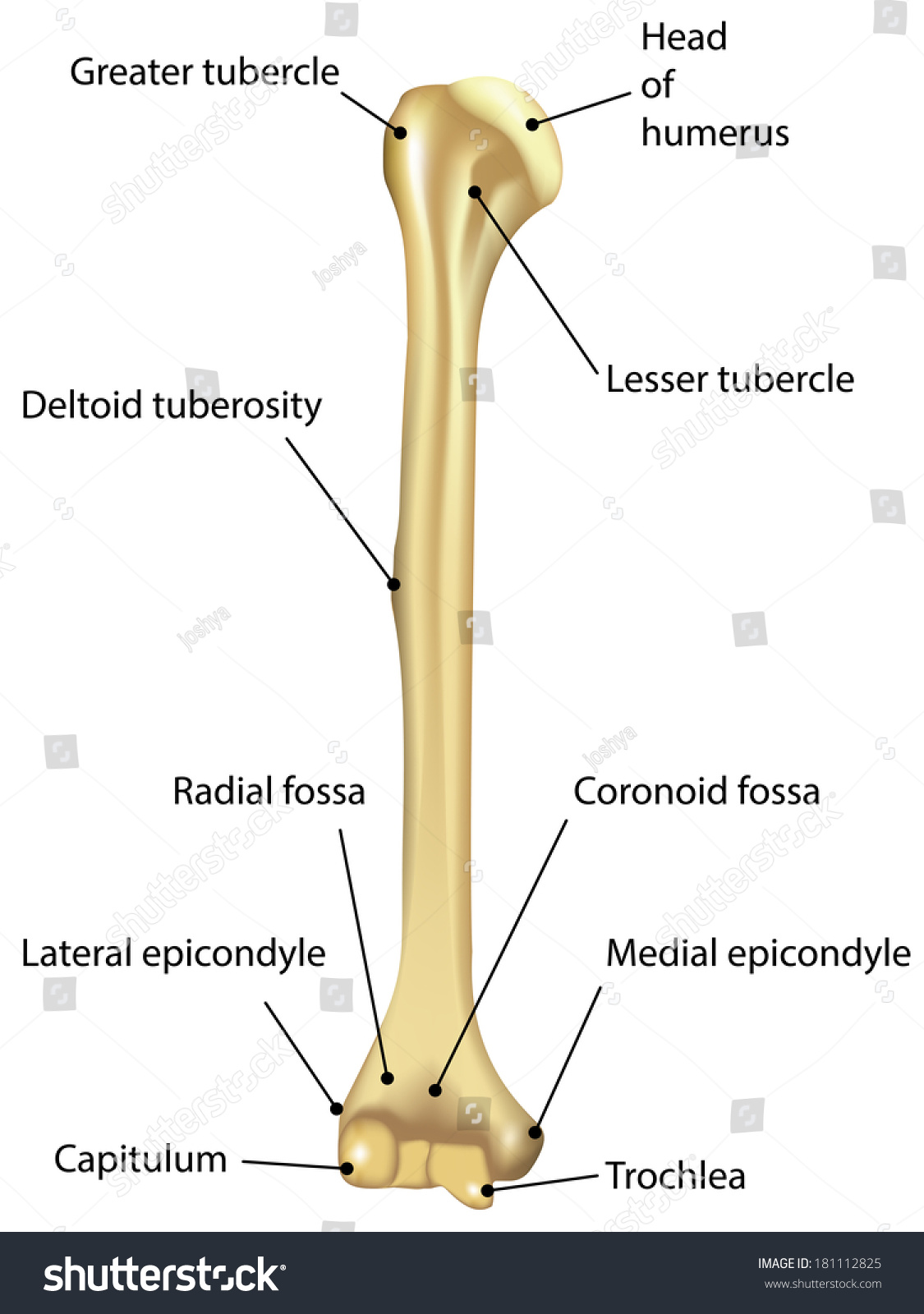
- Head of the Humerus: This is the rounded, smooth surface that articulates with the glenoid cavity of the scapula.
- Anatomical Neck: This is a slight constriction just below the head of the humerus.
- Greater Tubercle: This is a large, round protuberance on the sidelong side of the humerus, which serves as an attachment site for the rotator cuff muscles.
- Lesser Tubercle: This is a smaller prominence on the median side of the humerus, which also serves as an attachment site for the rotator cuff muscles.
- Intertubercular Groove: This is a deep groove that runs between the greater and lesser tubercles, supply a passage for the tendon of the long head of the biceps brachii muscle.
Shaft of the Humerus
The shaft, or body, of the humerus is relatively smooth and cylindrical, with a few notable humerus bone markings. These include:
- Deltoid Tuberosity: This is a rough, V shaped area on the sidelong side of the shaft, which serves as an attachment site for the deltoid muscle.
- Radial Groove: This is a shallow groove on the posterior surface of the shaft, which provides a passage for the radial nerve and the profunda brachii artery.
- Nutrient Foramen: This is a pocket-size open on the anterior surface of the shaft, through which blood vessels enter the bone to supply it with nutrients.
Distal Humerus Bone Markings
The distal end of the humerus features respective significant humerus bone markings that are all-important for interpret the bone s part and the muscles that attach to it. These markings include:
- Lateral Epicondyle: This is a outstanding bony process on the sidelong side of the distal humerus, which serves as an attachment site for the extensor muscles of the forearm.
- Medial Epicondyle: This is a prominent bony process on the median side of the distal humerus, which serves as an attachment site for the flexor muscles of the forearm.
- Trochlea: This is a smooth, pulley forge surface on the median side of the distal humerus, which articulates with the trochlear notch of the ulna to form the elbow joint.
- Capitulum: This is a smooth, round surface on the lateral side of the distal humerus, which articulates with the head of the radius to form the elbow joint.
- Coronoid Fossa: This is a shallow depression on the anterior surface of the distal humerus, which accommodates the coronoid process of the ulna during flexion of the elbow.
- Olecranon Fossa: This is a deep depression on the behind surface of the distal humerus, which accommodates the olecranon procedure of the ulna during extension of the elbow.
Clinical Significance of Humerus Bone Markings
Understanding the humerus bone markings is crucial for diagnosing and handle assorted injuries and conditions regard the humerus. for representative:
- Fractures: Fractures of the humerus can occur at several points along the bone, and cognition of the humerus bone markings can help identify the specific location and type of cracking.
- Dislocations: Dislocations of the shoulder or elbow joint can induce damage to the humerus bone markings, and see these markings can aid in name and treating these injuries.
- Muscle and Tendon Injuries: Injuries to the muscles and tendons that attach to the humerus bone markings can cause pain and limited range of motion. Knowledge of these markings can help place the specific muscles or tendons involved and guide treatment.
Note: The humerus bone markings are also important for surgical procedures involving the humerus, such as joint replacements or shift repairs. Surgeons must have a thorough understanding of these markings to ensure proper placement of implants and to avoid damaging nearby structures.
Imaging Techniques for Visualizing Humerus Bone Markings
Several imaging techniques can be used to visualize the humerus bone markings and diagnose injuries or conditions impact the humerus. These techniques include:
- X rays: X rays are commonly used to visualise the humerus and its humerus bone markings. They can help identify fractures, dislocations, and other abnormalities.
- Computed Tomography (CT) Scans: CT scans provide detail cross sectional images of the humerus and its humerus bone markings. They are useful for diagnose complex fractures and plan surgical procedures.
- Magnetic Resonance Imaging (MRI): MRI scans provide detailed images of the soft tissues circumvent the humerus, as easily as the bone itself. They are utilitarian for diagnosing muscle and tendon injuries, as good as other conditions affecting the humerus bone markings.
Common Injuries and Conditions Affecting the Humerus
Several injuries and conditions can affect the humerus and its humerus bone markings. Some of the most mutual include:
- Fractures: Fractures of the humerus can occur at various points along the bone, including the proximal, shaft, and distal regions. Common types of humerus fractures include:
| Type of Fracture | Description |
|---|---|
| Proximal Humerus Fracture | A fracture that occurs near the head of the humerus, often affect the greater or lesser tubercles. |
| Humeral Shaft Fracture | A fracture that occurs along the shaft of the humerus, often induce by direct trauma or a fall. |
| Distal Humerus Fracture | A faulting that occurs near the distal end of the humerus, frequently involve the lateral or medial epicondyles. |
- Dislocations: Dislocations of the shoulder or elbow joint can cause damage to the humerus bone markings and beleaguer tissues. Common types of dislocations include:
| Type of Dislocation | Description |
|---|---|
| Shoulder Dislocation | A dislocation that occurs when the head of the humerus is forced out of the glenoid caries of the scapula. |
| Elbow Dislocation | A disruption that occurs when the distal end of the humerus is pressure out of alignment with the bones of the forearm. |
- Muscle and Tendon Injuries: Injuries to the muscles and tendons that attach to the humerus bone markings can stimulate pain and limited range of motion. Common types of muscle and tendon injuries include:
| Type of Injury | Description |
|---|---|
| Rotator Cuff Tear | An injury that occurs when one or more of the rotator cuff tendons are torn, often involving the greater or lesser tubercles of the humerus. |
| Biceps Tendonitis | An injury that occurs when the tendon of the long head of the biceps brachii muscle becomes inflamed, much involving the intertubercular groove of the humerus. |
Note: Treatment for injuries and conditions involve the humerus and its humerus bone markings may include rest, ice, compression, elevation (RICE), physical therapy, medication, or surgery, depending on the severity of the injury.
Conclusion
The humerus is a complex bone with numerous humerus bone markings that play crucial roles in the movement and constancy of the amphetamine limb. Understanding these markings is essential for diagnose and handle diverse injuries and conditions affecting the humerus. By familiarize themselves with the anatomy and clinical significance of the humerus bone markings, medical professionals, anatomists, and students can gain a deeper discernment for the function and construction of this important bone.
Related Terms:
- humerus bone markings quiz
- femur bone markings
- humerus bone markings chart
- radius bone markings
- humerus bone unlabeled
- humerus bone markings labeled